组会讲课人员:赵月月
Identifying Extracellular Vesicle Populations Fromsingle Cells
从单个细胞中识别细胞外囊泡群
主讲人:赵月月
Proceedings of the National Academy of Sciences of the United States of America |September 2021| DOI: 10.1073/pnas.2106630118
Abstract:
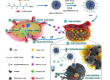
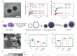
Extracellular vesicles (EVs) are constantly secreted from both eukaryotic and prokaryotic cells. EVs, including those referred to as exosomes, may have an impact on cell signaling and an incidence in diseased cells. In this manuscript, a platform to capture, quantify, and phenotypically classify the EVs secreted from single cells is introduced. Microfluidic chambers of about 300 pL are employed to trap and isolate individual cells. The EVs secreted within these chambers are then captured by surface-immobilized monoclonal antibodies (mAbs), irrespective of their intracellular origin. Immunostaining against both plasma membrane and cytosolic proteins was combined with highly sensitive, multicolor total internal reflection fluorescence microscopy to characterize the immobilized vesicles. The data analysis of high-resolution images allowed the assignment of each detected EV to one of 15 unique populations and demonstrated the presence of highly heterogeneous phenotypes even at the single-cell level. The analysis also revealed that each mAb isolates phenotypically different EVs and that more vesicles were effectively immobilized when CD63 was targeted instead of CD81. Finally, we demonstrate how a heterogeneous suppression in the secreted vesicles is obtained when the enzyme neutral sphingomyelinase is inhibited.
.
摘要:
细胞外囊泡(ev)是由真核细胞和原核细胞不断分泌的。ev,包括那些被称为外泌体的,可能对细胞信号传导和病变细胞的发病率产生影响。在这篇手稿中,介绍了一个捕获、量化和表型分类从单个细胞分泌的ev的平台。采用约300pL的微流控室来捕获和分离单个细胞。在这些腔室内分泌的ev然后被表面固定的单克隆抗体(mab)捕获,而不管它们的细胞内来源如何。对质膜和胞质蛋白的免疫染色结合高敏感度、多色全内反射荧光显微镜来表征固定的囊泡。对高分辨率图像的数据分析允许将每个检测到的EV分配到15个独特群体中的一个,并证明即使在单细胞水平上也存在高度异质性的表型。分析还显示,每个单抗分离物具有表型不同的ev,当靶向CD63而不是CD81时,更多的囊泡被有效固定。最后,我们演示了当酶中性的鞘磷脂酶被抑制时,在分泌的囊泡中如何获得异质性抑制。